Lethal bird flu virus

Although H5N1, the avian influenza virus, darling of a scare-mongering media is lethal. No doubt about it. It’s just that at this point in human history, this virus is not in a human transmissable form, thankfully.

Now, a new study of patients who became infected with H5N1 in Vietnam has revealed clues as to why the virus is so lethal.

Menno de Jong and colleagues compared viral levels and effects on the immune system in one group of patients infected with H5N1 and another group infected with two types of human flu virus. The patients with H5N1 infection had much greater viral load in the throat than the patients infected with the human virus; markers of viral load were highest in the H5N1 patients who died. Virus could also frequently be detected in the blood of H5N1 patients but only in those who died.

The authors found that H5N1 virus at high levels triggers the release of inflammatory cytokines and that levels of these compounds is associated with a higher viral load. Fatal H5N1 infection was also associated with a loss of white blood cells in the peripheral blood.

The authors posit that H5N1 replicates much faster than human flu virus and that the high levels of the virus trigger an overwhelming inflammatory response that contributes to lung dysfunction and eventually death. They report their results in Nature Medicine today.

An important point that should be made is that the lower virulence of human flu is the mechanism by which this virus has remained endemic in humans and infects millions year in year out. If it were highly virulent, like H5N1, it would kill too many hosts to be persistent year in year out. As a dead host cannot infect another in the same way as a living one in direct contact. It is not necessarily true that H5N1 will mutate immediately into such a persistent viral sub-strain. The likelihood is that it will mutate into a fast-infecting form initially, but such a form will remove hosts from the ecosystem too quickly to remain endemic in the way that common human flu is. Viz, we don’t see the 1918 flu today because it died out.

Sun, Sun, Sun

Girl SunbathingThere has been a lot of discussion over the summer as to whether we should all be getting a bit more sun to boost cancer-fighting vitamin D levels. That argument coupled with revelations that suntan creams might themselves boost the risk of skin cancer all fly in the face of the contrary view that we should be staying in the shade.
One thing the sun worshippers and those of tan-free skin agree on – getting sun burn is no fun. Now, researchers in England (A country fabled for its sunny climes) are giving volunteers a tan in a bid to find a treatment for sunburn. Anna Nicolaou of the University of Bradford, in the renowned sunspot of the North of England, is examining the biological mechanisms underlying sunburn and why it particularly affects people who don’t get a “good” tan.

The research team hopes to discover whether melanocytes that do not actively produce melanin discharge inflammatory mediators, including pro-inflammatory hormones called prostaglandins, which cause the redness, irritation and swelling of the skin that is observed in sunburn. They also hope to discover why people who tan easily are less likely to develop sunburn, contrasting to pale-skinned people who tend to sunburn easier.

You can find out more via the Bradford U press site.

Meanwhile, at the other end of the beach.

A new light-activated ingredient that mops up damaging iron could help reduce the effects of sunburn, according to research published in the Journal of Investigative Dermatology by UK scientists.

‘When skin is exposed to high doses of sunlight, such as when you are sunbathing, a massive amount of free iron is released in skin cells,’ explains Pourzand, ‘This free iron can act as catalysts for the generation of more harmful free radicals that cause severe cell damage.”

The net effect of mopping up iron released as the skin burns, is to reduce inflammation and pain, which are exacerbated by iron, and to prevent the build up of free radicals, which have been linked to an increased risk of skin cancer.

The researchers, Charareh Pourzand of the University of Bath and James Dowden, now at Nottingham University, are currently testing prototypes of the ingredient in the lab using three-dimensional human skin cultures and anticipate trialling the ingredient with human volunteers in the next two to three years.

Find out more at the Bath U press site

Crystallising thoughts on bird flu

UK researchers have determined the X-ray structure of a key protein of the avian influenza virus that reveals a peculiarity that might prove the Achilles’ heel of bird flu and allow new potent drugs against the disease that could stave off a flu pandemic.

H5N1 virus is named for the particular haemagglutinin (H) and neuraminidase (N) proteins that are found on the viral coat. Haemagglutinin allows the viral particles to bind to and infect host cells, while neuraminidase helps the virus to escape infected cells and attack new ones. There are 16 H’s and 9 N’s in known variants on the influenza type A virus.

It is the neuraminidases that are the target for drugs such as oseltamivir (Tamiflu) and zanamivir (Relenza). The trouble is, N1 is not the exact target for which they were designed. Rather the design of these drugs was based on neuraminidases N2 and N9 found in other influenza type A strains. H5N1 itself resides in a genetically distinct group. Structural information from this group could be critical to producing drugs that are resistant to emerging resistance in influenza.

Read the full story in my latest news round up at spectroscopynow.com

Nervous analysis

Chemical communication between nerve cells underlies brain functions, such as the control of sensory and motor processing, memory and learning. Monitoring neurotransmitter secretion is crucial to studying and understanding how the brain works. Various research teams have endeavoured to develop techniques for monitoring neurotransmitter release at the single-cell level, according to researchers in The Netherlands only invasive methods such as electrophysiological techniques and fluorescent probe labelling have provided adequate temporal and spatial sensitivity. These limits suffer from being limited to specific analytes.

Now, the researchers have turned to a specialist spectroscopic technique to develop a new approach to monitoring neurotransmitters that is non-invasive and side-steps the issue of analyte limitations.

Read the complete story in SpectroscopyNOW.com

Premature diagnostics

An MRI scan can help in predicting future developmental outcomes of pre-term infants, according to US researchers. The scan reveals abnormalities no picked up by cranial ultrasound and can be used to predict problems that would become apparent by age two years.

Terrie Inder of Washington University School of Medicine in St Louis working with colleagues in New Zealand and Australia have discovered that MRI can reveal brain abnormalities in very premature babies born at 30 weeks or less. They monitored 167 pre-term infants from birth to age two years having graded these abnormalities to see if there is a correlation with any severe delays in cognitive and psychomotor development, cerebral palsy, or hearing or visual impairments in the two-year olds.

Read on…

Sunscreen and skin damage

When I first wrote about the doubts scientists were raising concerning sunscreens in Chem & Industry magazine some time in the early 1990s, it seemed that the findings would simply confuse consumers and cause a storm among manufacturers. Unfortunately, that doesn’t seem to be the case, sun worshippers carried on frying themselves, slapping on only meagre amounts of purportedly protective cream, partly out of laziness and partly because it is just so expensive and manufacturers continued to sell their products by the bucket load. Then research emerged that showed lack of sun exposure not only risks rickets but could increase the chances of you getting internal cancers, as opposed to the much feared skin cancer. As if consumers were not confused enough.

Now, US researchers have shown that applying too little suncream can actually turn the UV-absorbing chemicals against you.

When skin is exposed to sunlight, ultraviolet radiation (UV) is absorbed by skin molecules that then can generate harmful compounds, called reactive oxygen species or ROS, which are highly reactive molecules that can cause “oxidative damage.” For example, ROS can react with cellular components like cell walls, lipid membranes, mitochondria and DNA, leading to skin damage and increasing the visible signs of aging. The link with skin cancer itself is actually not so clear cut as some lobbyists claim.

However, when sunscreen is applied to the skin the UV filters in the sunscreen, reduce the amount of UV radiation penetrating the skin. Over time, though, the filters themselves are absorbed by the skin leaving the surface vulnerable to UV once more. The UV filters (octylmethoxycinnamate, benzophenone-3 and octocrylene) widely used in sunscreens themselves generate reactive oxygen species (ROS) in skin when exposed to ultraviolet radiation, augmenting the ROS that is naturally produced. [These new results are similar to those on which I reported in C&I ten years ago, DB]

Kerry Hanson of the University of California Riverside and colleagues now report that these three UV filters only work well if sunscreen is reapplied frequently to prevent ultraviolet radiation from reaching these filters. Without reapplication, these compounds could be just as harmful as not using sunscreen at all.

The team will publish their findings in a forthcoming issue of Free Radical Biology & Medicine.

“Sunscreens do an excellent job protecting against sunburn when used correctly,” said Hanson, who works in the laboratory of Christopher Bardeen, an assistant professor of chemistry at UCR. “This means using a sunscreen with a high sun protection factor and applying it uniformly on the skin. Our data show, however, that if coverage at the skin surface is low, the UV filters in sunscreens that have penetrated into the epidermis can potentially do more harm than good. More advanced sunscreens that ensure that the UV-filters stay on the skin surface are needed; such filters would reduce the level of UV-induced ROS. Another solution may be to mix the UV-filters with antioxidants since antioxidants have been shown to reduce UV-induced ROS levels in the skin.”

“For now, the best advice is to use sunscreens and re-apply them often — the Skin Cancer Foundation recommends every two hours, and especially after sweating or swimming, which can wash away sunscreen — to reduce the amount of UV radiation from getting through to filters that have penetrated the skin,” Bardeen said. “This, in turn, would reduce ROS generation.”

But, having read this please also read my write-up on how to sunbathe safely

Statins, heart attacks, and a protection racket

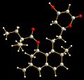
Structure of Simvastatin

Long-term treatment with statins help lower cholesterol and reduce heart attack and stroke incidence. But, it seems, GPs have not been prescribing enough as yet another benefit is now being touted in new research published today in the British Journal of Pharmacology. This latest study suggests this action only needs a few days of treatment, is independent of cholesterol lowering and helps the heart to recover following a heart attack.

Jing-Lin ZHAO and colleagues at the Fuwai Heart Hospital, Beijing, China, have demonstrate decreased ‘no-reflow’ and better preservation of ventricular function in animals treated for only two days with a statin called simvastatin (structure shown). They also identify the mechanism underlying this cardioprotective action, showing that the observed beneficial effects of simavastatin are abolished by blocking the KATP channel in mitochondria.

Following a heart attack, blood does not flow properly into vessels in the heart, even once they are re-opened. Increasing the blood flow in these vessels would decrease the area known as ‘no-reflow’ and restore more of the ventricular function. ZHAO’s results suggest that taking statins long-term not only reduces the likelihood of a heart attack, but could also give the heart a chance to recover from a heart attack by decreasing the damage done to heart muscle.

Of course, if those heart attack patients had been on statins in the first place…

Cellulite and weight loss

Cellulite weight lossCellulite is of growing concern to a huge number of women and many consider different ways of reducing it including bariatric surgery or a tummy tuck liposuction procedure, diets and other methods, while others really couldn’t care less about the superficial dimpling of their subcutaneous fat. Moreover, it provides yet another example of the medicalisation of a perfectly harmless “condition” as exemplified by the description of “sufferers” as patients.

But, recent research into weight loss and its effects on so-called cellulite is making a big splash. Losing weight can reduce the appearance of cellulite for some women, according to research undertaken by plastic surgeon John Kitzmiller and colleagues. But, here’s the rub, weight loss could make the condition much worse for others.

About 85% of women (that’s 8.5 women in every ten ;-) are affected by cellulite, Kitzmiller claims, “Cellulite is not specific to overweight people, but excess weight may worsen the condition. We found that weight loss in overweight patients improved the appearance of cellulite, but for a few, it actually worsened the condition.”

He examined 29 women [not the biggest most statistically significant sample] who enrolled in medically supervised weight loss programs including low-fat meals, liquid diets, medication, and bariatric surgery. Seventeen patients experienced an improvement in the appearance of their cellulite, while 9 worsened. The average weight loss was 30.5 pounds (range 2.3 — 102 pounds).

Those women who lost most weight and reduced their thigh fat too saw the greatest reduction in cellulite. These patients had a significantly higher starting body mass index (BMI) and had more severe cellulite on average. “Patients” whose cellulite worsened started with a significantly lower BMI, lost smaller amounts of weight, and had no change in percentage of thigh fat.

“There is no answer for completely eliminating cellulite, however, it appears the more weight one loses, the better its appearance,” explains Kitzmiller. “Although the appearance of cellulite diminished for the majority of patients, weight loss did not totally eradicate the condition. The dimples appear to be permanent features that lessen in depth as the pounds come off.”

Needless to say the cosmetic industry and plastic surgeons the world over will be rubbing their hands with glee at Kitzmiller’s revelations. Meanwhile, those women who should be reducing their weight for genuine health reasons who are looking for another reason to procrastinate may find the study most useful in providing them with another reason to put off that new fitness regime and low-cal diet.

Bird flu reaches the US

…but it’s not the deadly strain of avian influenza (high-pathogenicity avian influenza, HPAI H5N1) that has featured in endless media speculation over the last couple of years. At a time, when the Thai authorities have announced several new cases of bird flu in their country, scientists in the US have detected the low-pathogenicity (LPAI) bird flu in wild swans near the banks of Lake Erie.

Ron DeHaven, administrator of USDA’s Animal and Plant Health Inspection Service, told the Associated Press that “We do not believe this virus represents a risk to human health.” Preliminary testing showed the presence of the H5 and the N1 sub-types in the infected Michigan birds, but the USDA explains that these are more probably present because of LPAI, which is also known as American H5N1, as opposed to Asian H5N1.

Evidence of this putative low pathogenicity avian influenza (LPAI) has been found on two occasions in wild birds in the United States. In 1975 and 1986, it was detected in wild ducks. These detections occurred as part of routine sampling. LPAI H5N1 has also been detected in Canada, most recently in 2005.

Asian H5N1 has killed at least 140 people but is yet to mutate into a form that is readily transmissable between humans. Indeed, it seems the only likely way you catch this strain is through very close proximity to infected birds (i.e. sharing living quarters) or by coming into contact with feces from an infected bird.

More on the Michigan bird flu story…

Bird flu drugs

In the latest issue of The Alchemist on ChemWeb.com I provide a round-up of the week’s chemistry news, of particular importance could be news that UK and Australian researchers may have found a new way to approach drug design for bird flu viruses that precludes drug resistance.

A new drug to fight bird flu that should be able to side-step the emergence of viral resistance is being developed by Andrew Watts of the University of Bath, UK and Jennifer McKimm-Breschkin of CSIRO Australia. Both Tamiflu and Relenza, the two drugs currently being stockpiled by governments in preparation for a global outbreak of bird flu, are inherently susceptible to resistance because of the way they work. Although the new drug acts on the same target as these treatments, the enzyme neuraminidase, it targets a specific region of the enzyme that essential to its function. If this region mutates the virus would no longer be viable, so that resistance cannot emerge.

Read about this and more in this week’s Alchemist.