Salmonella Shows its Mettle

Salmonella bacteria use RNA to assess and adjust magnesium levels, according to researchers at the Washington University School of Medicine in St. Louis. Eduardo Groisman and colleagues at WUSTL have added a new gene to the bacterium via a mechanism known as the riboswitch.

Riboswitches were first identified in 2002 and sense when a protein is needed and stop the creation of the protein if it isn’t. A riboswitch, does not rely on anything binding to DNA; instead, the switch is incorporated into messages for construction of proteins. These messages are protein-building instructions copied from DNA into strands of RNA. The riboswitch is a sensor within the RNA that can twist it into different configurations that block or facilitate the production of the protein encoded in the message.

Previously identified riboswitches respond to organic compounds such as nucleotides and sugars. The Salmonella riboswitch, reported on Friday in Cell, responds to magnesium ions, key elements in the stability of cell membranes and reactants in an energy-making process that fuels most cells.

“Magnesium ions are essential to the stability of several different critical processes and structures in the cell, so there has to be a fairly intricate set of regulators to maintain consistent levels of it,” says senior investigator Groisman, “To approach such a complex system, we study it in a simpler organism, the Salmonella bacterium.”

Groisman and his colleagues uncovered the magnesium riboswitch while they were investigating the MgtA gene, which is controlled by the major regulator of Salmonella virulence, the phoP/phoQ system. The MgtA gene codes for a protein that can transport magnesium across the bacterium’s cell membrane. Groisman’s group showed 10 years ago that the phoP/phoQ system controls when Salmonella makes MgtA.

You can read more about the work at the WUSTL site.

Tuberculosis Waste Disposal Defeats Immune System

The first detailed structure of a crucial protein-cleaving component, the proteasome, commonly known as the cellular waste disposal unit, of the tuberculosis bacterium has been obtained by US researchers. The existence of a proteasome in this microbe, only hinted at previously, could offer new targets for drug research to treat the disease.

Read on…

For more science news with a spectral angle visit my spectroscopy news page.

Pinning Down Alzheimer Clue

US researchers have used a powerful spectroscopic technique to demonstrate that an enzyme previously shown to protect brain cells from the characteristic fibrous tangles associated with Alzheimer’s disease also helps inhibit formation of the amyloid peptide plaques (APPs) seen in this disease. The team examined the relationship between APPs and the enzyme prolyl isomerase, Pin1.

The full story is now available on the spectroscopy site – SpectroscopyNOW.com

You can access my other spectroscopy news stories via this Sciencebase page.

Blood Group Test

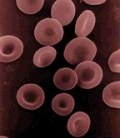
red-blood-cellsA, B, AB, or O?

A blood type (also called a blood group) is a classification of blood based on the presence or absence of inherited antigenic substances on the surface of red blood cells (RBCs). These antigens may be proteins, carbohydrates, glycoproteins, or glycolipids, depending on the blood group system, and some of these antigens are also present on the surface of other types of cells of various tissues. Several of these red blood cell surface antigens, that stem from one allele (or very closely linked genes), collectively form a blood group system.

The ABO system is the most important blood group system in human blood transfusion. The associated anti-A antibodies and anti-B antibodies are usually “Immunoglobulin M”, abbreviated IgM, antibodies. ABO IgM antibodies are produced in the first years of life by sensitization to environmental substances such as food, bacteria and viruses. The “O” in ABO is often called “0” (zero/null) in other languages.

A quite literally vital question when a blood transfusion is required and normally blood type is determined using an antibody and optical examination. However, Austrian researchers at the University of Vienna have developed a novel approach that is much simpler and side-steps expensive antibodies. Their technique is based on the blood-type-specific adsorption of red blood cells (erythrocytes) on a plastic surface “embossed” on the molecular scale.

Production of the analytical chips needed for this method is a simple and inexpensive process: quartz microbalances (tiny piezoelectric quartz crystals) are coated with a wafer-thin film of polyurethane. Erythrocytes of a specific blood type in liquid are placed on a slide and stick to its surface, forming the embossing “stamp”. The polymer is cured to harden it and the cells washed off. The ebmossed plastic surface now contains a large number of tiny impressions with indentations shaped like the antigens on the surface of the blood cells.

If a sample of blood is then placed on the chip, the erythrocytes will preferentially settle into those impressions with a matching shape. The resulting increase in mass is measured with the incredibly sensitive quartz microbalance.

The shape and size of the erythrocytes are the same for all blood types, so how can they be differentiated by these indentations? ‘The outer form is not the deciding factor,’ says team leader Franz Dickert, ‘instead, it is the differences in the surfaces of the different blood types.’ There are sugar-like molecular fragments on the surface of the cells that differentiate the blood types.

‘Despite a noticeable cross-sensitivity for the other blood types, determination of the blood type by the embossed plastic films is unambiguous,’ says Dickert, ‘because the strongest sensor signal comes from the microbalance that carries the impressions corresponding to the blood type of the sample.’

Dickert and colleagues publish details of their technique in Angew Chem, 2006, 45, 2626-2629

The Dynamic Duo of Biology

Researchers have modified a popular system for protein labelling and modification to reduce the risk of unwanted cross-reactions and so make it more accurate and effective.

With incredible specificity and powerful affinity for each other, the protein streptavidin and its small-molecule target biotin are truly the ‘Dynamic Duo’ of biological research, the researchers explain, and a perennial favourite for use in the design of biochemical experimental techniques. For example, one can easily subject biotin-linked proteins to highly specific labelling with streptavidin-linked fluorophores. Nonetheless, there is an important limitation to the system-streptavidin naturally forms tetramers (assemblies of four protein molecules) that bind up to four molecules of biotin, creating the potential for unexpected cross-linking of biotinylated targets. Efforts to engineer monomeric streptavidin variants have generally resulted in diminished biotin affinity.

Now, Alice Ting and colleagues at Massachusetts Institute of Technology, Cambridge, have developed an alternative approach that involves engineering ‘dead’ streptavidin variants that can bind to each other but not to biotin. By combining the two types of streptavidin monomers in the proper proportions and isolating tetramers that consist of three dead subunits and one active subunit, they obtain streptavidin complexes that are functionally monomeric and bind only one molecule of biotin.

They have demonstrated that the hybrid tetramers retain normal affinity for biotin but induce far less ‘clumping’ of biotinylated targets relative to wild-type streptavidin tetramers. This approach also offers the possibility of building divalent and trivalent tetramers. According to Kai Johnsson of the École Polytechnique Fédérale de Lausanne, Lausanne, Switzerland, “the existing plentitude of applications of the streptavidin-biotin interaction provides an enormous playground for streptavidins with reduced but defined valencies.”

More details can be found in April’s Nature Methods.

Metabolic Reaction

Portuguese researchers have developed a technique for classifying genomic-wide metabolic reactions, which they suggest will open up a new approach to diversity analysis of metabolic reactions and comparison of metabolic pathways as well as being generally compatible with the conventional “EC” classification of enzymes.

You can read my complete story on this at spectroscopnow.com

Unleaded Babies

Exposure to environmental poisons is suspected of disturbing the secondary sex ratio, i.e. the difference in numbers of girls and boys born as opposed to the ratio of girls and boys conceived. Indeed, several countries, including Canada, Denmark, England and Wales, Germany, The Netherlands, and the USA, have seen the secondary sex ratio shift during the last hundred years or so that the number of girls among live-births has risen significantly. One of the more significant declines is seen in Mexico, although some countries, Ireland, in particular, have seen the reverse, with more boys.

You can read the complete story in my news story on SpectroscopyNOW.com

Keeping Pace with Genetic Variation

A gene associated with disease might vary from a healthy gene in one individual DNA base pair – a so-called point mutation. Investigating point mutations and diagnosing genetic disease would benefit from a simple, cost-effective and rapid sequencing technique.

Now, Japanese researchers have developed a new approach for a miniaturized system that detects small differences in DNA sequences with high sensitivity. In contrast to other methods, this technique works without labeling the bases and exploits a field effect transistor (FET) to detect changes in the charge on DNA molecules.

According to Toshiya Sakata and Yuji Miyahara multiple FETs can feel electrical fields and react to changes by changing the current that flows through their conducting channels. The researchers loaded the surface of an FET with short, single-stranded pieces of DNA. These probes are the exact counterparts to the sequence at the beginning of the DNA segment being investigated. If a sample containing the target DNA comes into contact with the surface, the target DNA binds to the probes. The polymerase chain reaction (PCR) is then used to reconstruct the complete target DNA strand. Cleverly, the team do not use all four DNA building blocks at once but dip the FET into four different solutions, each containing only one of the building blocks, one after the other. After each dip, the electrical characteristics of the FET are measured. If and only if a component has been added to the end of the chain, a change is registered. This occurs because each building block brings with it a negative charge, which changes the electrical field on the surface of the FET. In this way, DNA chains of a length up to about ten components can be precisely sequenced. Missing, extra, or changed nucleotides can be rapidly and unambiguously identified.

You can read more details in Angew Chem Int Edn, 2006, 45, 2225

Lazarus Rats

Mammalian paleobiologists must be a cynical lot. How else to explain their presumably ironic use of the term Lazarus Effect in piecing together the very evidence for evolution that must by its nature preclude much of what is discussed in the same context as Lazarus? Moreover, this particular piece of evidence is one that plugs a gap in the fossil record and helps bring continuity to the theory of evolution, something that is often considered a fatal flaw by those who’d dispossess it. You could say they’ve brought it back to life…

Anyway, a new type of rodent discovered last year in Laos is a survivor of a group believed to
have been extinct for 11 million years, according to a new study published today in Science. Mary Dawson and colleagues compared skeletal remains of the squirrel-like animal with those of a little understood extinct group of Southeast Asian rodents and confirmed that it is actually a living member of this long-gone family. When the new species was discovered in early 2005, it drew wide acclaim because it was thought to be a member of an entirely new family of living mammals. Instead, according to the researchers, the rodent represents a striking example of the ‘Lazarus effect’ in which an organism suddenly reappears after a long gap in its fossil record. That such a phenomenon has only rarely been documented among mammals and other vertebrates shows that Southeast Asia’s prehistoric ‘zoo’ can offer invaluable insights regarding past and present
biodiversity, the researchers write.

Missing Bio Link

The missing piece in the biochemistry of haem (heme) is reported in this week’s spectroscopyNOW. Japanese researchers have used x-ray diffraction to determine the crystal structure of the enzyme human indoleamine 2,3-dioxygenase (IDO). This crucial enzyme splits the pyrrole ring of the amino acid L-tryptophan and incorporates both atoms of a molecule of oxygen, an essential step in dozens of metabolic reactions. The discovery could have implications for other studies involving this enzyme and medical problems with which it is associated.