Spray-on sex could usher in an age of McNookie, according to an article in The Observer on Sunday. PT-141 is billed as libido in an atomiser, says the paper, and could finally offer women the chance to turn on their sexual desire as and when they need it.
“A dose of PT-141 results, in most cases, in a stirring in the loins in as little as 15 minutes,” reports Julian Dibbell, “Women, according to one set of results, feel ‘genital warmth, tingling and throbbing’, not to mention ‘a strong desire to have sex’.”
So, what is PT-141?
It’s an odourless and colourless synthetic chemical that you inhale deeply through a small, white plastic inhaler. The compound, produced by Palatin Technologies and currently undergoing regulatory assessment, is a melanocortin-based therapy that seems to work directly on the brain rather than simply stimulating the loins as is the case with Viagra.
The drug’s market name is Bremelanotide and it’s a word you’re sure to see a lot more of in the near future as spammers start offering it and variations on the theme (watch out for Bremelan0tide in your email subject lines). Its mode of action is poorly understood, but unlike thousands of years of rhino horn, tiger dick, and oysters this one definitely seems to work.
“It’s not merely allowing a sexual response to take place more easily,” Michael Perelman, co-director of the Human Sexuality Program at New York Presbyterian Hospital and a sexual-medicine adviser on the PT-141 trials explains, “It may be having an effect, literally, on how we think and feel.” What is known is that it acts on at least one of five known melanocortin receptor subtypes in the brain. These are chemical receptors that regulate a diverse array of functions including sexual arousal, appetite, energy maintenance and inflammation. The company is working on drugs to affect these receptors and so help with sexual dysfunction, obesity and cachexia (disease-related muscle loss).
One has to wonder though whether the advent of drugs like PT-141 is simply another example of the kind of drug mongering I discussed a few years ago my Alchemist column on ChemWeb. There now seem to be dozens more “diseases” than there ever were. Is it just coincidence that problems that were not considered problems are now categorised as syndromes and disorders have emerged at just the time when the patents are expiring on the billion dollar staples of the pharmaceutical industry? Maybe, maybe not. Some of those disorders, such as restless legs syndrome, may sound like spurious ailments invented by a desperate industry, but they certainly aren’t for those who suffer from them. Even shyness and anxiety might one day succumb to drugs, and why not? If these disorders are debilitating then who are we to suggest that sufferers have no right to a treatment that “cures” them?
Sometimes there is no relief for a whole range of problems without pharmaceutical intervention. Whether or not we should allow people the option to control their sexual desire chemically is a moot point, the fact is humans have used all kinds of methods to loosen up a libido for millennia, and will continue to do so whether that’s with a regulated drug or something else. Palatin seems simply to be hoping to take a slice out of the action.
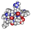
Anyway, for those interested in such things Palatin gives the basic structure of its product as a short cyclic peptide with the sequence – Ac-Nle-cyclo[Asp-His-D-Phe-Arg-Trp-Lys]-OH. It’s currently in Phase III clinical trials, so it shouldn’t be too long before those spams start arriving.